Can a healthy baby have a thick nuchal fold?
Nuchal folds are common in normal babies. A thick nuchal fold in a healthy baby can be caused by a number of factors including the position of the baby, inaccurate measurements, and typically the normal size differs for every baby. The use of NT thickness as a screening tool for fetal abnormalities is still debatable.
What is the nuchal skin fold at 20 weeks?
All babies have a small pad of tissue at the back of the neck; this is called the nuchal fold (NF). The nuchal fold normally measures less than 6mm at 20 weeks.
What is the normal range for nuchal fold?
What is the NT scan normal range? At 11 weeks, the skin fold measures up to 2 mm. At 13 weeks and 6 days, it can be up to 2.8 mm. If your baby’s measurements are within this range, they have a low chance of having Down syndrome or other genetic disorders.
What is the nuchal thickness for trisomy 21?
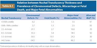
In the majority of fetuses with trisomy 21, the nuchal translucency thickness was <4.5 mm, while with trisomies 13 or 18 it was 4.5-8.4 mm, and in those with Turner syndrome, it was 8.5 mm or more 9.
How thick is the nuchal fold at 24 weeks?
Nuchal fold thickness increased with GA in a linear manner from 3.13 ± 0.68 mm (mean ± SD) at 16 weeks to 5.08 ± 0.76 mm at 24 weeks. The 95th percentile measurement at 24 weeks remained less than 6 mm.
What is an increased nuchal fold at 22 weeks?
An increased nuchal fold could be a sign for trisomy 21, but most often it is a normal variation in the baby’s development. Your health-care practitioner might offer you a referral to a genetics/MFM centre to discuss your results, and options for more testing.
When to stop measuring nuchal folds?
Nuchal fold thickness of >6 mm is abnormal on a routine morphology ultrasound performed at 18-22 weeks. The nuchal fold is known to increase throughout the second trimester in a normal pregnancy, and may be measured during a broader window of 14 and 24 weeks when required.
Can nuchal fold measurement be wrong?
False positives can be common in nuchal translucency tests, particularly due to their nature as a prenatal screening, which signifies that it cannot diagnose a condition. This is why many physicians will typically combine an NT screening with a blood test.
Do boys have thicker nuchal fold?
Male fetuses had a higher mean NFT as compared to female fetuses in the entire cohort (3.7 mm in males versus 3.5 mm in females, p < 0.0001, 95% CI 0.3-0.1).
What is a good nuchal result?
The cut-off for the nuchal translucency measurement is 3.5 mm. If your measurement is less than 3.5 mm, this is considered “normal”. If your measurement is 3.5 mm or more, this is considered “increased”. The ultrasound also looks at the baby’s body parts, like the heart and the brain.
What is a high risk nuchal result?
If you have a ‘high risk result’ it means it is more likely that there is an abnormality present. If your test returns a ‘high-risk’ result, your doctor can refer you for further testing. You may decide to have a diagnostic test, which can give you a definite result.
What if nuchal thickness is 1.3 mm?
If the fetal ultrasound measures nuchal translucency below 1.3mm, the risk of Down syndrome will be very low. If the fetal ultrasound measures nuchal translucency higher than 3mm, there is a risk of Down syndrome.
What is a thickened NT range?
The presence of a thickened NT (≥3.5 or ≥3.0 mm) is associated with common and uncommon aneuploidies and with a wide variety of genetic syndromes and structural anomalies, even when the karyotype is normal. Alamillo C.M.
What is the NT size for Down syndrome?
This baby with an NT of 6mm has a high chance of Down’s syndrome, as well as other chromosomal and heart conditions (NHS England 2023, Simpson 2021). It’s rare for babies to have as much fluid as this. Up to 14 weeks, your baby’s NT measurement usually increases as they grow.
How accurate is nuchal thickness?
NT screenings alone can detect about 70% of trisomy 21, or Down syndrome cases. Many healthcare providers combine a normal NT ultrasound with blood screenings. The accuracy of predicting conditions increases to about 95% when combined with first-trimester blood tests.
Do all Down syndrome babies have thick nuchal fold?
All unborn babies have some fluid at the back of their neck. In a baby with Down syndrome or other genetic disorders, there is more fluid than normal. This makes the space look thicker. A blood test of the mother is also done.
What is an enlarged nuchal fold?
Increased measurement of the nuchal fold (≥ 6 mm from 14 weeks to 22 weeks of gestational age) is considered a soft marker for chromosomal aneuplodies, as well as for structural defects in the fetus, most commonly cardiac defects. We also describe the outcome of fetuses with increased second trimester nuchal folds.
What is normal nuchal thickness?
The distribution of the NT thickness for CRL has been reported in many studies. The median NT thicknesses has been reported to be 1.2-1.9 mm, 1.22-2.10 mm, and 1.19-1.73 mm for a CRL between 45 mm and 80 mm in Japan, Korea, and Brazil, respectively.
What should nuchal fold be at 23 weeks?
Nuchal fold thickness of >6 mm is abnormal on a routine morphology ultrasound performed at 18-22 weeks. The nuchal fold is known to increase throughout the second trimester in a normal pregnancy, and may be measured during a broader window of 14 and 24 weeks when required.
Can a baby with high NT be normal?
Fetuses with a nuchal translucency index lower than 1.3mm have a very low risk of Down syndrome. Nuchal translucency lower than 2.5mm is considered a safe range. When the nuchal translucency is high from 2.5mm to 6mm, doctors need to add the results of other indicators to draw conclusions.
What if my baby has increased nuchal translucency?
When there is an increased NT measurement, there is a higher chance for the baby to have: a chromosome difference, such as trisomy 21 (Down syndrome) and trisomy 18 (Edwards syndrome).
Is a 4mm nuchal fold normal?
In fetuses with nuchal translucency of 4 mm or more and normal karyotype, there was a high association with other defects and the prognosis was often poor, whereas the translucency resolved for those with 3 mm and the pregnancy outcome was usually normal.
How often is the NT measurement wrong?
The accuracy rate of Nuchal Translucency (NT) ultrasound screening in identifying babies’ risk factors for chromosomal abnormalities is 70- to 75- percent when used as a standalone risk assessment with a five-percent false-positive rate.
Can nasal bone develop after 20 weeks?
We have established the nasal bone length in South Indian fetuses at 16–26 weeks of gestation and there is progressive increase in the fifth percentile of nasal bone length with advancing gestational age. Hence, gestational age should be considered while defining hypoplasia of the nasal bone.
Why is nuchal fold thickness at 20 weeks?
Second trimester thickened nuchal fold has a high specificity for aneuploidy. ACOG and SMFM define an abnormal nuchal fold as ≥ 6mm between 15 and 20 weeks of gestation. It is the most powerful second trimester sonographic marker for Trisomy 21.
Can a thick nuchal translucency go away?
It is known that most cases of increased NT observed in the first trimester disappear as pregnancy progresses. We therefore investigated the frequency of fetal abnormalities in such cases. The average NT thickness in cases of increased NT that later disappeared was 4.1 ± 1.4 (mm).
What is abnormal nuchal measurement?
Due to this the NT measurement may considered abnormal when it is above 3.0 mm, or above the 99th percentile for the gestational age. In pooled data from 30 studies, NT screening alone has a sensitivity for trisomy 21 of 77% with a 6% false-positive rate.
Can a thick nuchal translucency be normal?
When the NT is thickened, above what is generally considered normal for the baby at that gestational age (usually more than 3-3,5mm), there is an increased risk for the baby of having a chromosomal anomaly (Down syndrome or others).
Can a baby with a normal nuchal fold have Down syndrome?
Normal Results A normal amount of fluid in the back of the neck during ultrasound means it is very unlikely your baby has Down syndrome or another genetic disorder. Nuchal translucency measurement increases with gestational age. This is the period between conception and birth.
What does it mean when a fetus has a thick neck?
All unborn babies have some fluid at the back of their neck. In a baby with Down syndrome or other genetic disorders, there is more fluid than normal. This makes the space look thicker. A blood test of the mother is also done.
What causes nuchal thickening?
The proposed etiology of increased nuchal thickness is the result of hydrops or lymphatic obstruction.
How thick is a nuchal fold?
Does nuchal fold thickness increase with gestational age?
When is a nuchal fold normal?
What is a fetal nuchal fold?
You’re 23 weeks pregnant, and your doctor just mentioned something called “nuchal fold thickness.” Maybe you’re a little confused, maybe a little worried. Don’t stress! We’re here to break down what this measurement means and why it’s important.
What is Nuchal Fold Thickness?
The nuchal fold is a small fold of skin at the back of a baby’s neck. It’s basically like a little bit of extra skin there. The nuchal fold thickness is the measurement of this fold. It’s measured during an ultrasound, usually between 11 and 14 weeks of pregnancy, but sometimes it can be measured later, like at 23 weeks.
Why Measure the Nuchal Fold at 23 Weeks?
You might be thinking, “Why measure it at 23 weeks? Isn’t that a bit late?” You’re right, the ideal time to measure nuchal fold thickness is between 11 and 14 weeks. But sometimes, it’s not possible to get an accurate measurement during that window. This can happen for a few reasons:
Baby’s Position: The baby might be in a position that makes it difficult to get a clear image of the nuchal fold.
Mom’s BMI: A higher BMI can make it harder to get a good view of the baby.
Technical Issues: Sometimes, there are issues with the ultrasound machine or the equipment that prevent a clear measurement.
If you didn’t get the measurement between 11 and 14 weeks, your doctor might recommend measuring the nuchal fold at 23 weeks. It’s important to understand that measuring at 23 weeks is not as accurate as measuring earlier in the pregnancy, but it can still be helpful.
What Does the Nuchal Fold Measurement Tell Us?
The nuchal fold measurement is used to assess the risk of certain chromosomal abnormalities in your baby. Chromosomal abnormalities are changes in the number or structure of chromosomes. These changes can lead to a variety of health problems, including Down syndrome, trisomy 18, and trisomy 13.
A thick nuchal fold is often associated with these chromosomal abnormalities. However, it’s important to remember that a thick nuchal fold doesn’t necessarily mean your baby has a chromosomal abnormality. Many babies with thick nuchal folds are perfectly healthy.
How is Nuchal Fold Thickness Measured?
The nuchal fold is measured using an ultrasound. The ultrasound technician will use a specialized probe to get a clear image of your baby’s neck. They will then measure the thickness of the nuchal fold at its widest point.
Nuchal Fold Thickness at 23 Weeks: Interpreting the Results
The meaning of your nuchal fold measurement at 23 weeks will depend on a few factors:
Your Baby’s Gestational Age: The measurement is interpreted differently depending on how far along you are in your pregnancy.
Your Family History: Your doctor will consider if there’s a family history of chromosomal abnormalities.
Other Risk Factors: Other factors, like your age and previous pregnancies, can also be considered.
If your nuchal fold measurement is above the normal range, your doctor will likely recommend further testing, such as amniocentesis or chorionic villus sampling (CVS). These tests can provide more definitive information about your baby’s health.
What if the Nuchal Fold Measurement is Normal?
If your nuchal fold measurement is within the normal range, it’s a good sign! This doesn’t guarantee that your baby is 100% healthy, but it does lower the risk of certain chromosomal abnormalities. Your doctor will likely continue to monitor your pregnancy with regular ultrasounds.
Nuchal Fold Thickness at 23 Weeks: FAQs
1. Is it normal to have a nuchal fold thickness measured at 23 weeks?
Yes, it’s not uncommon for the nuchal fold to be measured at 23 weeks if it wasn’t possible to measure it earlier in the pregnancy.
2. What is the normal range for nuchal fold thickness at 23 weeks?
The normal range for nuchal fold thickness varies depending on the baby’s gestational age. Your doctor will be able to tell you what the normal range is for your baby’s specific age.
3. What if my nuchal fold measurement is above the normal range at 23 weeks?
If your nuchal fold measurement is above the normal range, your doctor will likely recommend further testing to determine the cause. This doesn’t necessarily mean your baby has a chromosomal abnormality, but it’s important to get a clearer picture.
4. Does a normal nuchal fold measurement at 23 weeks mean my baby is healthy?
A normal nuchal fold measurement at 23 weeks is a good sign, but it doesn’t guarantee that your baby is 100% healthy. There are other factors that can contribute to a healthy pregnancy.
5. What should I do if I have concerns about my nuchal fold measurement?
If you have any concerns about your nuchal fold measurement, it’s best to talk to your doctor. They can help you understand the results and discuss your options.
Remember, a nuchal fold measurement is just one piece of information about your baby’s health. It’s important to keep the big picture in mind and talk to your doctor about any questions or concerns you may have.
See more here: What Is The Nuchal Skin Fold At 20 Weeks? | Nuchal Fold Thickness At 23 Weeks
Prenatal diagnosis and pregnancy outcome analysis of thickened
The measurement of nuchal fold (NF) thickness during the second trimester is considered to be one of the most sensitive and specific isolated ultrasound National Center for Biotechnology Information
260: Second trimester nuchal fold thickness and
Increased measurement of the nuchal fold (≥ 6 mm from 14 weeks to 22 weeks of gestational age) is considered a soft marker for chromosomal aneuplodies, as well as for structural defects in the fetus, most American Journal of Obstetrics & Gynecology
Measurement of nuchal skin fold thickness in the second
Comparison of nuchal skin fold thickness (NFT) in a normal 20-week fetus in the breech and transverse presentations. (a) A sonogram in the breech presentation demonstrates an increased NFT Obstetrics and Gynecology
Ultrasonographic Soft Markers of Aneuploidy in
Nuchal Fold Thickening. Nuchal edema in the second trimester between 15 and 23 weeks is known as the nuchal fold. Nuchal thickening was the first of the nonstructural markers… Medscape
Impact of Gestational Age on Nuchal Fold Thickness in
Nuchal fold thickness increased with GA in a linear manner from 3.13 ± 0.68 mm (mean ± SD) at 16 weeks to 5.08 ± 0.76 mm at 24 weeks. The 95th percentile Wiley Online Library
Prenatal ultrasound: Increased nuchal fold (2nd trimester)
The nuchal fold is a normal fold of skin at the back of a baby’s neck. This can be measured between 15 to 20 weeks in pregnancy as part of a routine prenatal ultrasound. The My Doctor Online
A pictorial guide for the second trimester ultrasound – PMC
Less than 6 mm is considered normal up to 22 weeks. When measuring the nuchal fold angling the probe to place the falx at ~15° to horizontal may provide a National Center for Biotechnology Information
Nuchal Translucency Screening – What to Expect
What is a nuchal translucency test and what does it measure? A nuchal translucency screening, or NT screening, is a specialized routine ultrasound performed at the end of the first What to Expect
Nuchal scan – Wikipedia
The nuchal fold thickness is considered normal if under 5mm between 16 and 18 weeks gestation and under 6mm between 18 and 24 weeks gestation. An increased thickness Wikipedia
See more new information: curtislovellmusic.com
Nt (Nuchal Translucency) And Nf (Nuchal Fold) U/S Measurements. The Difference And Upper Limits.
Fetus Ultrasound, Thickened Nuchal Fold
Nuchal Translucency (Nt) Thickness Measurement: For Early Fetal Scan Conference 2019
Nuchal Translucency (Nt)
What Is A Normal Nuchal Translucency Measurement?-Dr. Geeta Komar
Thick Nuchal Fold Nt Scan | Positive Outcome
Increased Nuchal Fold Thickness + Dilated 4Th Ventricle
Diagnosis Of Down Syndrome
Link to this article: nuchal fold thickness at 23 weeks.

See more articles in the same category here: https://curtislovellmusic.com/category/what/