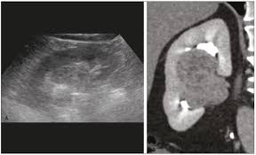
What are the ultrasound features of the column of Bertin?
The following are characteristic of a hypertrophied column of Bertin: It is a projection of cortex into the renal sinus (and therefore is isoechogenic with it). The sinus may engulf it in a clawlike fashion. The renal contour is smooth. Sonography is characteristic and obviates further investigation.
What are the columns of Bertin?
Columns of Bertin represent the extension of renal cortical tissue which separates the pyramids, and as such are normal structures. They become of radiographic importance when they are unusually enlarged and may be mistaken for a renal mass (renal pseudotumor).
What are the columns of Bellini?
The renal column (or Bertin column, or column of Bertin) is a medullary extension of the cortex in between the renal pyramids. Ducts of Bellini, also called Papillary ducts. The duct of Bellini represents the most distal portion of the collecting duct. It allows the cortex to be better anchored.
What is the function of the column of Bertini?
Columns of Bertini are also known as renal columns. Its main function is to make the cortex anchored in a better manner to the medullary region. It is made up of fibrous material and also has blood vessels and urinary tubes passing through them.
What are the main features of ultrasound?
Ultrasound imaging (sonography) uses high-frequency sound waves to view inside the body. Because ultrasound images are captured in real-time, they can also show movement of the body’s internal organs as well as blood flowing through the blood vessels.
What is a bertin?
: any of the masses of cortical tissue extending between the sides of the renal pyramids of the kidney as far as the renal pelvis.
What are the columns of Bertin in the kidneys of mammals formed as an extension of?
Therefore, the column of Bertini in the kidneys of mammals is formed as an extension of cortex in the medulla.
What does the name Bellini mean?
Bellini is a gender-neutral name of Italian origin, meaning “little beautiful one.” This endearingly sweet and loving name can serve as a perfect reminder for your little one wherever they go.
What does bellini mean in Italian slang?
Bellini originates from the word Bella meaning. beautiful in many languages. Bellini is also a pretty. chill person she gets a long with almost everybody …
What do the columns of Bertini divide the medulla into?
(ii) Medulla is divided into few renal pyramids. (iii) Pyramid projects into calyx. (iv) Inward extension of cortex between the pyramids is called renal column of Bertini. III.
What is the main function of a column?
Columns are frequently used to support beams or arches on which the upper parts of walls or ceilings rest. In architecture, “column” refers to such a structural element that also has certain proportional and decorative features.
What is the origin of the renal column of Bertini?
After French anatomist Exupere Joseph Bertin (1712–1781) described that renal cortical substance extends through pyramids in 1774, those cortical substances (septa) that separates the medullary pyramids and extends towards renal pelvis were named as columns of Bertin [1].
What are the 3 main types of ultrasound?
transrectal ultrasound, which provides images of the prostate gland. Doppler ultrasound, which monitors blood flow in the major arteries and veins. echocardiogram, which examines the heart. 3D ultrasound, which shows a three-dimensional picture of the inside of the body.
What are the components of ultrasound image?
A typical ultrasound imaging system uses a wide variety of transducers optimized for specific diagnostic applications. Each transducer is comprised of an array of piezoelectric transducer elements that transmit focused energy into the body and receive the resulting reflections.
What is the function of the column of Bertin?
Columns of Bertin represent the extension of renal cortical tissue which separates the pyramids, and as such are normal structures. They become of radiographic importance when they are unusually enlarged and may be mistaken for a renal mass (renal pseudotumor).
Does everyone have a column of Bertin?
Columns of Bertin represent the extension of renal cortical tissue which separates the pyramids and present in ~50% of the healthy population and in 20% are bilateral. They are usually located in the mid-portion of the kidney and are more commonly found on the left side (as in this case).
What is the hyperechoic column of Bertin?
A hypertrophied column of Bertin is one of the congenital causes of renal pseudo tumor. The columns of Bertin are normal structures seen in the renal cortical tissue. In 1744, French anatomist Exupere Joseph Bertin explained that the renal cortex extended in radial fashion surrounding the renal pyramids.
What are the renal columns called?
The renal columns, Bertin columns, or columns of Bertin, a.k.a. columns of Bertini are extensions of the renal cortex in between the renal pyramids. They allow the cortex to be better anchored. (Cortical extensions into the medullary space.)
Does the cortex extends in between the medullary pyramids as renal columns called columns of Bertini?
The cortex extends in between the medullary pyramids as renal columns called Columns of Bertin. The renal pyramids, renal columns and the cortex above it is known as the renal lobe.
What are the extensions of the kidneys?
Anatomical Position. The kidneys lie retroperitoneally (behind the peritoneum) in the abdomen, either side of the vertebral column. They typically extend from T12 to L3, although the right kidney is often situated slightly lower due to the presence of the liver. Each kidney is approximately three vertebrae in length.
What is a Bellini in English?
A Bellini is a cocktail made with Prosecco and peach purée or nectar. It originated in Venice, Italy. Pour peach purée into chilled glass, add sparkling wine.
What does Zygmunt mean?
With origins primarily in Poland but also linked to Germany, Zygmunt is a boy’s name meaning “conquering protection.” It’s derived from the name Sigmund, best known for Freud and can be shortened to a variety of nicknames, including Ziggy and Iggy. Perhaps Zygmunt is the perfect name for your little Starman.
Is Beatrice a Russian name?
Beatrice is a feminine name of Latin origin.
What are the ultrasound features of pyonephrosis?
Ultrasonographic findings suggestive of pyonephrosis include the presence of hydronephrosis in conjunction with hyperechoic debris in the collecting system (see the image below). The presence of debris and layering of low-amplitude echoes in the hydronephrotic kidney indicate pyonephrosis.
What are the sonographic features of acute focal nephritis?
We defined acute focal bacterial nephritis based on sonographic findings (focal hyper- or hypoechoic lesion with poor perfusion, irregular and poorly demarcated margins, possibly associated to significant renal enlargement) or CT findings (poorly demarcated areas with a wedge shape that are not enhanced after infusion …
What are the sonographic features of multicystic kidney disease?
The most useful ultrasonographic criteria for identifying multicystic kidney include: (a) the presence of interfaces between cysts (accurate in 100% of cases); (b) nonmedial location of the largest cyst (100%); (c) absence of an identifiable renal sinus (100%); (d) multiplicity of oval or round cysts that do not …
What is the sonographic feature of pyelonephritis?
In two patients with acute pyelonephritis, the ultrasonic findings consist- ed of a large swollen kidney with an increased anechoic corticomedullary area, with multi- ple scattered low-level echoes.
What are the sonographic characteristics of the column of Bertin?
What are the ultrasonographic features of hypertrophied columns of Bertin?
What does a Bertin column look like during intravenous urography?
What does a column of Bertin represent?
Let’s talk about ultrasound images and a specific feature you might see: the column of Bertin. It can be a bit confusing, especially if you’re new to the world of kidney imaging.
Think of it this way: your kidneys are like complex organs, with intricate internal structures. The column of Bertin is one such structure, and it can sometimes show up on ultrasound as a distinct feature.
What Exactly is the Column of Bertin?
Essentially, it’s a band of renal tissue, like a bridge of kidney tissue, that stretches inwards from the outer part of the kidney towards the center. Think of it as a kind of “bridge” within the kidney.
Now, you might be wondering, why is this important? Well, sometimes when we look at an ultrasound image, this column of Bertin might look a little suspicious, maybe even like a tumor. That’s why it’s crucial for us to understand what it is and how to distinguish it from other, potentially more concerning, abnormalities.
Why does the Column of Bertin Show Up on Ultrasound?
The column of Bertin is a natural part of the kidney, but it doesn’t always show up clearly on ultrasound. Often, it’s obscured by the surrounding kidney tissue. However, under certain conditions, it can become more prominent, making it visible on the ultrasound image.
Size: A larger-than-average column of Bertin is more likely to be seen on ultrasound.
Location: Its location within the kidney can also influence its visibility.
Patient Position: The way the patient is lying during the ultrasound scan can sometimes make it more prominent.
Differentiating the Column of Bertin from Potential Problems
Here’s where things get interesting. Sometimes, the column of Bertin can look remarkably similar to a tumor on ultrasound. That’s why it’s essential for radiologists to know what they’re looking at.
To tell them apart, we rely on a few key features:
Location: The column of Bertin is typically located centrally, often near the renal pelvis (the kidney’s “drainage system”).
Shape: It’s usually triangular or wedge-shaped, with its base facing the outer edge of the kidney.
Echogenicity: The column of Bertin usually has the same echogenicity (how the tissue reflects sound waves) as the surrounding kidney tissue.
When to be Concerned
While the column of Bertin is usually harmless, there are instances where it can raise concerns:
Unilateral: If it’s only present on one side of the kidney, it could potentially indicate a developmental abnormality.
Unusual Appearance: If it has an unusual shape, size, or echogenicity, it could be a sign of a more serious condition.
The Takeaway
The column of Bertin is a common feature on ultrasound images. While it can sometimes mimic a tumor, understanding its characteristics and location can help radiologists confidently identify it. Remember, it’s a natural part of the kidney, and in most cases, it’s nothing to worry about.
FAQs about the Column of Bertin
Q: Can the Column of Bertin Cause Pain?
A: Usually, the column of Bertin doesn’t cause any symptoms. It’s a normal anatomical variation and shouldn’t cause pain or discomfort.
Q: Is the Column of Bertin a Sign of Kidney Disease?
A: No, the column of Bertin itself is not a sign of kidney disease. It’s simply a normal anatomical feature of the kidney.
Q: Can the Column of Bertin Be Removed?
A: Removal of the column of Bertin is not typically necessary. It’s a normal part of the kidney and doesn’t require surgical intervention.
Q: What are the Potential Complications of the Column of Bertin?
A: The column of Bertin itself rarely causes complications. However, its appearance on ultrasound can sometimes lead to unnecessary investigations, especially if it’s misinterpreted as a tumor.
Q: Can the Column of Bertin Change Over Time?
A: The column of Bertin is a relatively stable feature. It’s unlikely to change significantly over time.
Q: How is the Column of Bertin Diagnosed?
A: The column of Bertin is usually diagnosed during an ultrasound examination. The radiologist will use the characteristics of the structure and its location to identify it.
Q: Should I be Worried if I Have a Column of Bertin?
A: In most cases, you shouldn’t be worried. It’s a normal anatomical variation. However, if you have any concerns, it’s always best to discuss them with your doctor.
Remember, the column of Bertin is a normal feature of the kidney that can sometimes cause confusion on ultrasound images. By understanding its characteristics and location, radiologists can accurately identify it and avoid unnecessary concerns.
See more here: What Are The Columns Of Bertin? | Column Of Bertin Ultrasound Images
Hypertrophic Columns of Bertin: Imaging Findings – PMC
Hypertrophic column of Bertin (HCB) may mimic renal mass and may lead to unnecessary nephrectomy in some conditions. In this case report we present a patient National Center for Biotechnology Information
Renal column of Bertin | Radiology Case | Radiopaedia.org
Ultrasound images of the right kidney in the long axis show a U-shaped protrusion of the renal cortex into the hilum. Case Discussion. Classical appearance of a Radiopaedia
Pictorial review: Renal ultrasound – Clinical Imaging
A prominent (or “hypertrophic”) column of Bertin can be mistaken for a renal tumor, leading to further imaging. Sonographic characteristics suggestive of the column of Bertin, though not Clinical Imaging
Imaging and Management of Incidental Renal Lesions – PMC
Hypertrophied column of Bertin. Coronal reformatted contrast-enhanced CT images in corticomedullary (a) and nephrographic phase (b) well demonstrate the National Center for Biotechnology Information
Hypertrophied column of Bertin | Radiology Case | Radiopaedia.org
On ultrasound the column splits the sinus echoes. CECT is often done to resolve the issue. A hypertrophied column shows enhancement that is the same as normal cortex. Radiopaedia
Congenital Anomalies of the Upper Urinary Tract: A
(b) US image in a 30-year-old woman shows a hypertrophied column of Bertin (*). (c) Coronal T2-weighted MR image in a 40-year-old man shows a dromedary hump (arrowhead) from the RSNA Publications Online
Hypertrophied Column of Bertin: A Mimicker of a Renal Mass
A contrast-enhanced computed tomography scan finalized the diagnosis of hypertrophied column of Bertin, as there was uniform uptake of contrast noted in the UroToday
Sonography of the hypertrophied column of Bertin | AJR
A prospective sonographic analysis of kidneys in 136 adults without clinical or radiologic evidence of renal disease revealed 22 cases of large columns of Bertin. Most were AJR
Hypertrophic columns of bertin: imaging findings – PubMed
Hypertrophic column of Bertin (HCB) may mimic renal mass and may lead to unnecessary nephrectomy in some conditions. In this case report we present a patient with HCB, PubMed
See more new information: curtislovellmusic.com
Hypertrophied Or Prominent Column Of Bertin, Ultrasound And Color Doppler Video
Columns Of Bertin Radiology #Radiology #Urology #Kidney
Prominent Column Of Bertin
Renal Columns Of Bertin Anatomy Named After People 🔊
Bertin’S Column
Ultrasound Imaging Of The Kidneys || Dr S Boopathy || Anatomy, Variations, Tips And Tricks On Usg
Sonography Renal Mass Case Studies
Ultrasound Of The Urinary Tract
Link to this article: column of bertin ultrasound images.

See more articles in the same category here: https://curtislovellmusic.com/category/what/